
What is VEGF
VEGF is a natural protein in the body that plays a key role in creating new blood vessels. Under normal conditions, VEGF is very important for healing wounds and forming blood supply in tissues that need more oxygen.
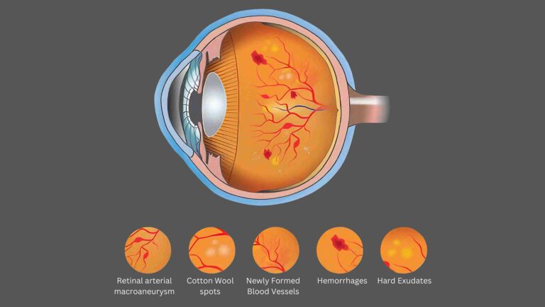
However, in the eye, too much VEGF can be harmful. When overproduced, it causes abnormal and fragile blood vessels to grow in the retina. These vessels often leak fluid or blood, leading to swelling and damage in the central part of the eye (the macula). This is what happens in diseases like:
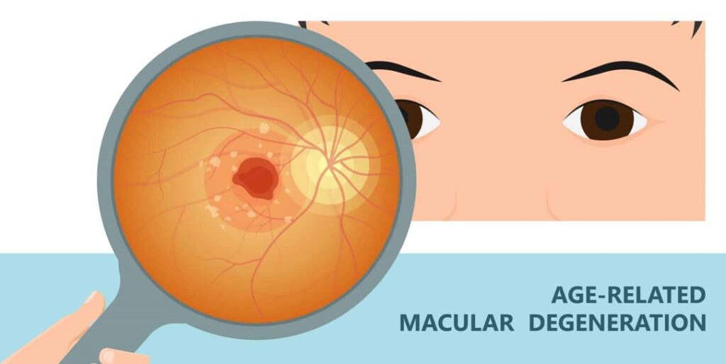
1. Wet Age-related Macular Degeneration (AMD)
2. Diabetic Retinopathy
3. Retinal Vein Occlusion
That’s why Anti-VEGF injections are used to block this protein, slow down or stop the abnormal growth of blood vessels, and protect vision. This method is often referred to as anti vegf injection treatment.
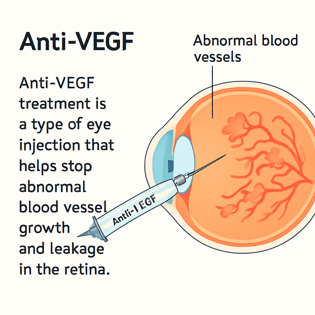
What is Anti-VEGF injection
Anti-VEGF injection (anti–vascular endothelial growth factor) treatment is a type of eye injection that helps stop abnormal blood vessel growth and leakage in the retina. It’s commonly used to treat vision-threatening conditions like diabetic macular edema (DME), age-related macular degeneration (AMD), and retinal vein occlusion (RVO). By blocking VEGF protein, Anti-VEGF injetion helps reduce swelling, prevent vision loss, and sometimes even improve eyesight.
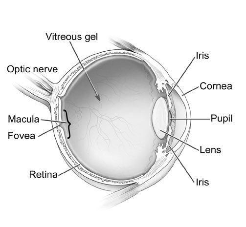
Anatomy
1. Outer (Fibrous)
(i) Sclera
(ii) Cornea
2. Middle (Vascular)
(i) Choroid
(ii) Iris
(iii) Ciliary Body
3. Inner: Retina
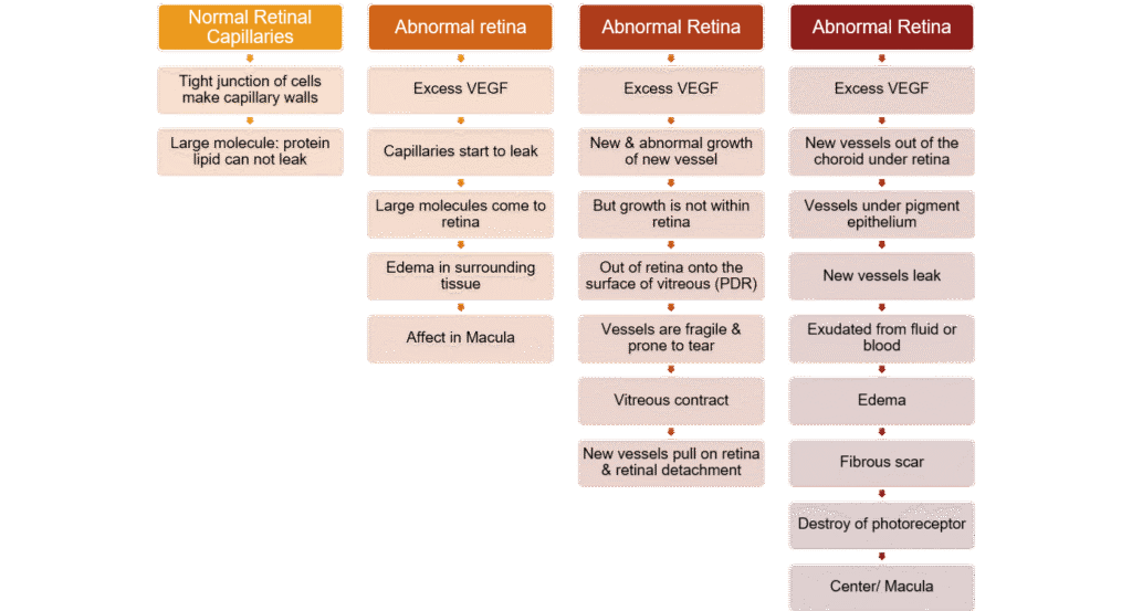
Type of VEGF
• In a healthy individual with a normally functioning retina, VEGF levels are typically regulated and maintained at an appropriate balance.
• VEGF is present at baseline levels to support the normal growth and maintenance of blood vessels in the retina, ensuring proper oxygen and nutrient supply to retinal tissues.
• In the absence of any pathological conditions, VEGF doesn’t cause excessive blood vessel growth or leakage.
• While VEGF is normally present to support healthy blood vessel growth in the retina, its dysregulation can lead to pathological neovascularization and leakage in retinal diseases.
• Managing VEGF levels through targeted treatments is an important strategy for addressing these conditions and preventing vision loss.
Type of Anti-VEGF injection
• Anti-VEGF injection medicine blocks VEGF, slowing the growth of blood vessels in the eye.
• This slows or stops damage from the abnormal blood vessels and slows down vision loss. Sometimes it can even improve vision.
• The effects of anti VEGF injection may be summarized as:
• Increased permeability of existing blood vessels, causing them to leak.
• Growth of abnormal new blood vessels, which may bleed or leak fluid and proteins.
In the eye, both can lead to retinal damage.
• Vascular endothelial growth factor (VEGF) is a protein produced by cells in your body. VEGF produces new blood vessels when the body needs them.
• Anti-VEGF injection agents, with the effects of reducing retinal neovascularization (RNV) and choroidal neovascularization (CNV), and inhibiting vascular permeability
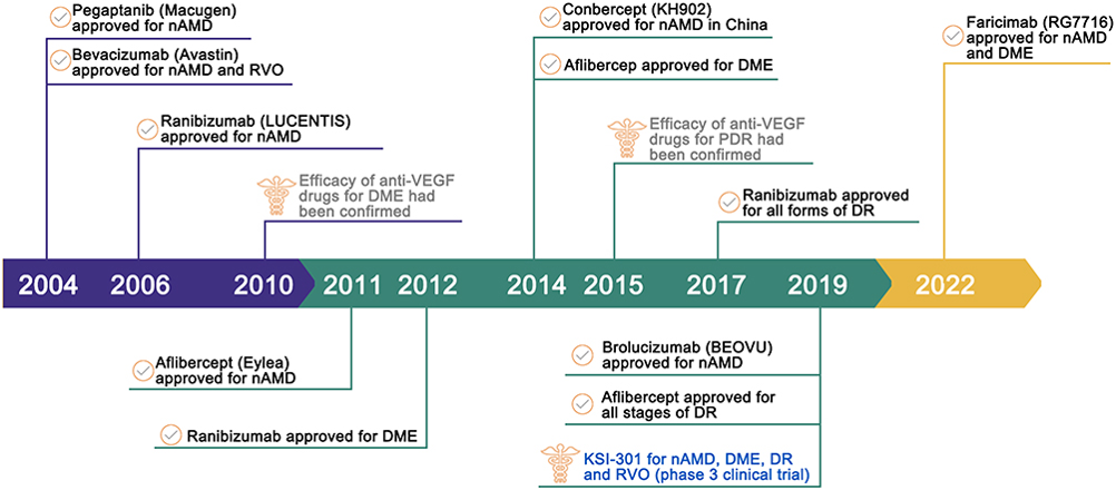
Anti VEGF injection approval & evolved
Present & Future of Anti-VEGF injection
In summary, anti vegf injection is a crucial intervention in managing various retinal conditions that threaten vision.
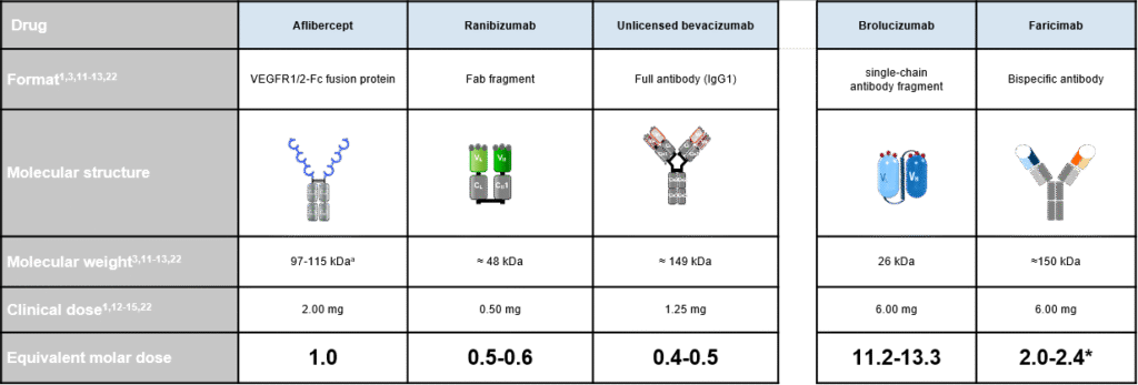
aMolecular weight expressed as a range to reflect glycosylation status. *Faricimab 6 mg is 4 times molar dose of 0.5 mg ranibizumab
CH, constant heavy; CL, constant light; Fab, fragment, antigen-binding; Fc, fragment crystallizable; IgG, immunoglobulin G; VEGFR, vascular endothelial growth factor receptor; VH, variable heavy; VL, variable light.
3. Holz FG, et al. Ophthalmology. 2016;123:1080-1089; 11. Avastin [package insert]. South San Francisco, CA: Genentech, Inc.2016; 12. Eylea [package insert]. Tarrytown, NY: Regeneron Pharmaceuticals, Inc. 2017; 13. Lucentis [package insert]. South San Francisco, CA: Genentech, Inc. 2017; 14. CATT Research Group. N Engl J Med. 2011;364:1897-1908; 15. IVAN Study Investigators. Ophthalmology. 2012;119:1399-1411; 22. Dugel PU, et al. Ophthalmology. 2017;124:1296-1304; 1. Sahni, J et al. Ophthalmology 2019;126:1155–70
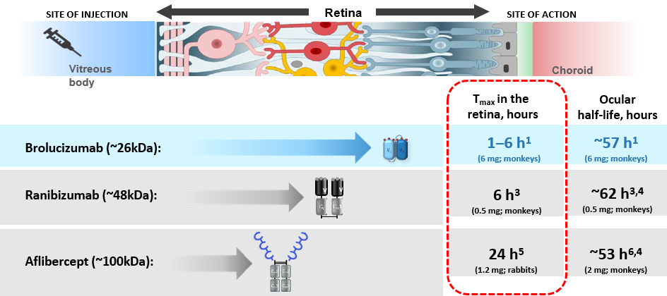
Brolucizumab penetrates the retina rapidly in preclinical studies
• Anti-VEGF injeciton need to penetrate the dense cellular network of the retina to inhibit the growth of new blood vessels in the RPE and choroid.
• All anti-VEGF injection remain in the eye for a similar length of time (ocular half-life), but the small size of brolucizumab may facilitate rapid and more effective penetration of the different retinal layers.
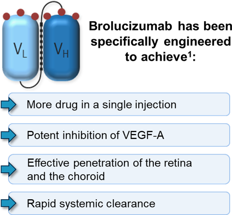
Brolucizumab – A novel anti-VEGF injection
Retinal Disease
DME, nAMD, BRVO, ROP, DME (Diabetic Macular Edema)
DR is a common, progressive microvascular complication of diabetes and is a precursor to PDR and DME
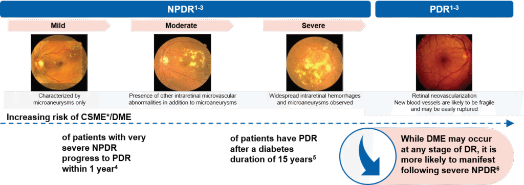
*CSME is a categorization used in most epidemiology studies to identify DME. While some clinicians point out distinctions, DME and CSME are considered synonymous; Fundus images taken from El-Bab MF, et al. Clin Ophthalmol. 2012;CSME, clinically significant macular edema; DME, diabetic macular edema; (NP/P) DR, (nonproliferative/proliferative) diabetic retinopathy. 1. Eshaq RS, et al. Pathophysiology 2017;24:229–41; 2. Coetting C. Modern Optometry, June 2019; 21-24; 3. ETDRS Study Group. Ophthalmology. 1991 May;98(5 Suppl):823-33; 4. Martinez-Zapata MJ, et al. Cochrane Database Syst Rev 2014;11:CD00872; 5. Fong DS, et al. Diabetes Care 2004;27:584–7; 6. Klein R, et al. Arch Ophthalmol 2001;119:547–53.
Accumulation of fluid in the layers of the macula
is an ubiquitous manifestation of retinal disease1
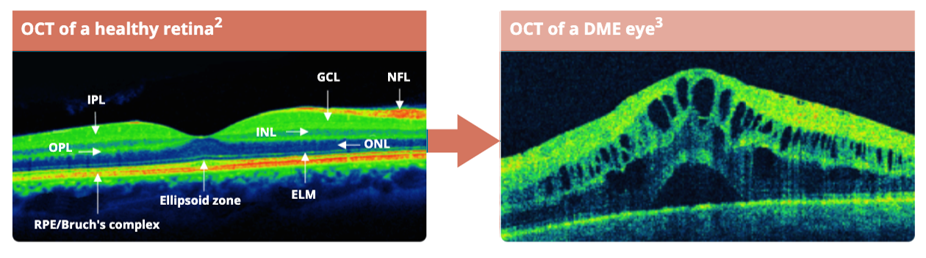
The interstitial spaces of
the retina are normally
maintained dry1
Retinal edema, is directly associated
with breakdown of the blood
retinal barrier and with
vision loss1
Figures reproduced with permission from publisher.
DME, diabetic macular edema; ELM, external limiting membrane; GCL, ganglion cell layer; INL, inner nuclear layer (bipolar cell); IPL, inner plexiform layer; NFL, nerve fiber layer; ONL, outer nuclear layer (photoreceptors); OPL, outer plexiform layer; RPE, retinal pigment epithelium
1. Cunha-Vaz J. Ophthalmologica. 2017;237:1–10. 2. Romero-Aroca P, et al. J Diabetes Res. 2016:2156273. 3. Eyeguru.org. How to read OCTs: 8 fundamental diseases. Available at: https://eyeguru.org/essentials/interpreting-octs/ [Accessed February 2021].
DME is characterized by abnormal accumulation of fluid in
the intraretinal and subretinal space in the macula, causing retinal thickening and leading to distortion of central vision1
In patients with diabetes, increased
blood sugar levels damage the retinal microcirculation which increases vascular permeability and results in leakage of fluid into the intraretinal layer2,3

Disruption of retinal architecture due
to fluid accumulation (macular edema)
leads to compromised visual function3,4
Representation of the macula region in a normal and a DME eye5
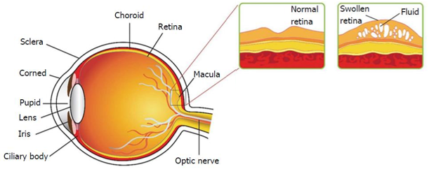
Figure reproduced with permission from publisher.
DME, diabetic macular edema
1. Chung YR, et al. J Diabetes Res. 2019:8164250. 2. Strain WD, et al. Diabetes Res Clin Pract. 2017;126:1–9. 3. Daruich A, et al. Prog Ret Eye Res. 2018;63:20–68.
4. Coughlin BA, et al. Vis Res. 2017;139:93–100. 5. Trinh HM, et al. World J Pharmacol. 2016;5:1–14.
DME is initiated by the breakdown of the inner BRB
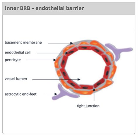
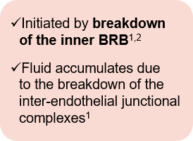
Figure reproduced with permission from publisher.
BRB, blood-retinal barrier; DME, diabetic macular edema; nAMD, neovascular age-related macular degeneration; RPE, retinal pigment epithelium
1. Cunha-Vaz J. Ophthalmologica. 2017;237:1–10. 2. Ragelle H, et al. J Ocul Pharmacol Ther. 2020;36:30–41.
DME treatment evolution over the past decades
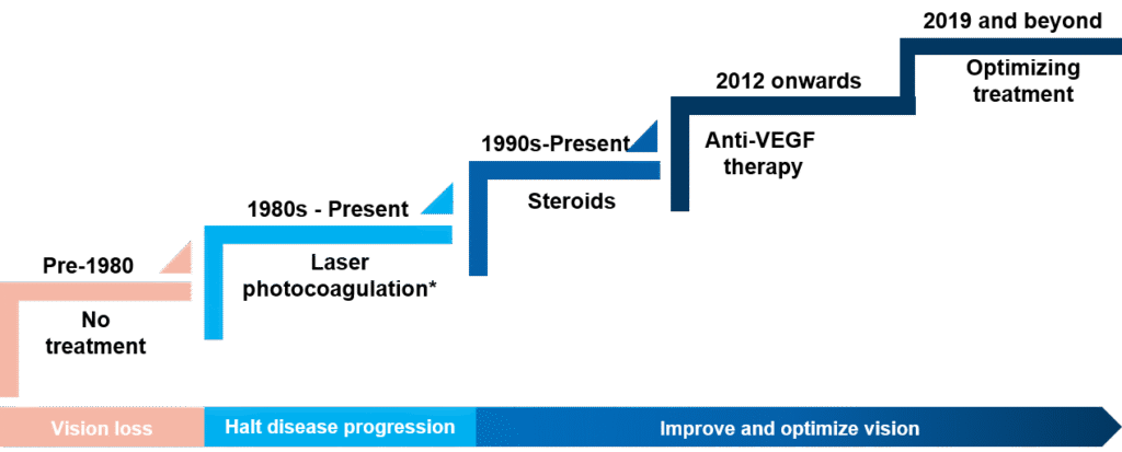
*Use has become limited; DME, diabetic macular edema; VEGF, vacular endothelial growth factor; 1. Schmidt-Erfurth, U et al, Ophthalmologica 2017;237:185-222
Resolving retinal fluid is essential for the treatment of nAMD patients
ANY FLUID is considered pathological in AMD
Disease progression: Fluid accumulation leads to a damaged retina and severe vision loss1
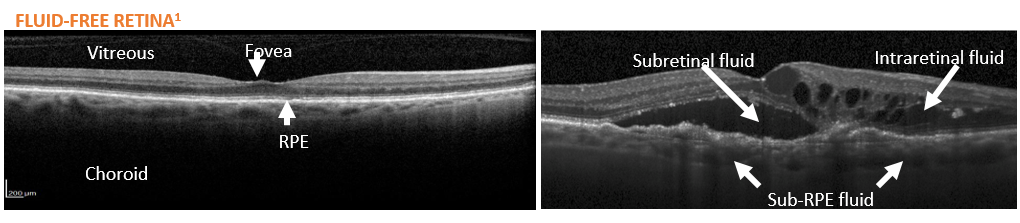
Healthy retina:
No fluid present
Drusen formation and VEGF overproduction
Fluid accumulation
Damaged retina and severe vision loss

All retinal fluid is considered a disease state and needs to be controlled with
appropriate treatment strategies to prevent irreversible vision loss.
1. Angiogenesis Foundation http://www.scienceofamd.org/resources/ [accessed Nov 2019]; 2. Novartis data on file; 3. Image reproduced with permission from Schmidt Erfurth U & Waldstein SM. Prog Ret Eye Res. 2016;50:1-24.
nAMD, neovascular age-related macular degeneration; RPE, retinal pigment epithelium
Neovascular AMD
- Wet, or exudative, AMD is diagnosed when choroidal neovascularization (CNV) is present in the eye.
- CNV is an in-growth of new vessels from the choriocapillaris into the sub-RPE space, via a break in Bruch’s membrane.
- Any disturbance of Bruch’s membrane, such as thickening or drusen, can weaken the membrane, increasing the likelihood that a break will occur and allow buds of CNV to invade the sub-RPE space.
- CNV vessels tend to extravasate serous fluid and blood into the retinal and sub-retinal space.
- The blood may resorb over time, dissect further under the retina, or even break through the retina and enter the vitreous space.
Therapeutic milestones for nAMD
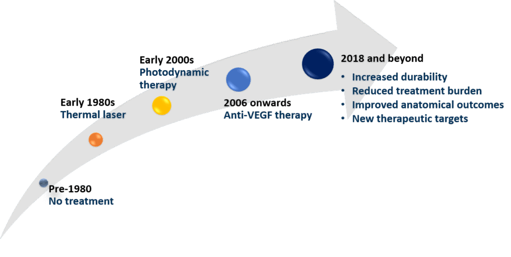

Anti-VEGF injection have completely changed the way doctors manage many serious eye diseases. Before this treatment, conditions like wet age-related macular degeneration or diabetic retinopathy often led to permanent and severe vision loss, leaving patients with very few options. Anti-VEGF injection therapy works by directly targeting the VEGF protein, which is the main trigger behind the growth of abnormal, leaky blood vessels in the retina. By blocking this process, the injections help reduce swelling, prevent bleeding, and protect the delicate retinal tissues that are essential for clear vision.
The biggest advantage is that, with regular treatment, patients can maintain their sight for many years, and in some cases, even regain vision that was already lost. This makes a huge difference in daily life allowing people to continue reading, working, or even just recognizing the faces of loved ones. Of course, it’s not a one-time cure; most patients need ongoing injections, sometimes for life. There may also be mild side effects like discomfort or temporary redness, and in rare cases, more serious complications.
Even with these limitations, Anti-VEGF injection are considered one of the greatest medical advances in eye care. They offer real hope where once there was none, giving patients the chance to live more independently and confidently, while preserving one of the most precious senses vision.
very good and informative article